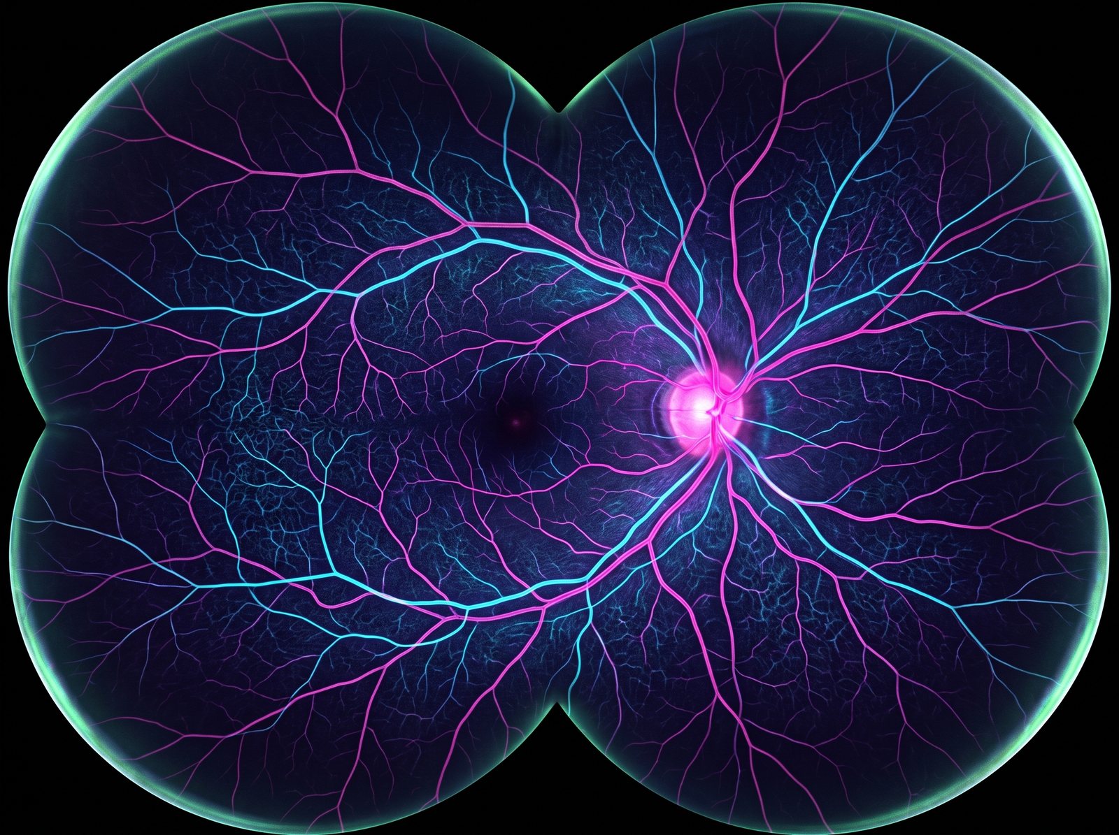
Cutting-edge technology in ophthalmology

We see your eye like never before.

Behind fifty years of eye surgery at Dr. P. Siva Reddy Eye Hospital sits an imaging and surgical arsenal that maps your eye to the micron — and reveals what a routine check never could.

0
Founded in Himayat Nagar — fifty years of eye surgery.
0
Generations of surgeons, tracing the history of cataract surgery itself.
0
Rating across 1,106 Google reviews.
0
Advanced diagnostic & surgical systems under one roof.
Why the machine matters// The arsenal

The eye is the only organ a doctor can examine while it looks back — and the only one where a fraction of a millimetre changes everything.

Three generations of surgeons built this hospital. What they have added for you is a suite of imaging and surgical systems that see, map and treat the eye at a scale the human eye simply cannot. Here is what each one means for the person in the chair.

ZEISS CLARUS 500 · ULTRA-WIDEFIELD
// Retinal imaging

Your whole retina, in a single frame.

An ordinary exam sees the centre of your retina. This captures almost all of it — out to the far edges where diabetes, tears and early disease hide — in one ultra-widefield image, often without dilating drops. Trouble is caught where it begins, not after it has already cost you sight.

Powered by ZEISS CLARUS 500
Field of viewUp to ~200°
CaptureSingle shot, drop-free
FindsDiabetic retina, tears
ZEISS CIRRUS 6000 · OCT ANGIOGRAPHY
// Depth & blood flow

A map of the blood flow inside your eye — no dye, no needle.

Without a single drop of dye, this scanner maps the living blood flow of your retina — every branch down to the finest capillary — and slices the tissue into cross-sections thinner than a human hair. Diabetic damage, macular disease and glaucoma surface years before you would ever feel them.

Powered by ZEISS CIRRUS 6000 OCT-A
AngiographyDye-free, needle-free
FieldUp to 12×12 mm widefield
DetectsDiabetic · AMD · glaucoma
ALCON VERION · IMAGE-GUIDED
// Cataract surgery

Your surgery, planned onto your eye before the first touch.

Before surgery your eye is measured and a digital plan is built for it. In the operating microscope that plan is projected straight onto your eye, guiding every incision and the exact placement of your new lens. The cataract itself is removed through an opening about two millimetres wide.

Alcon VERION guidance + LEGION phaco
PlanningDigital eye-mapping
Incision~2 mm micro-incision
LensesMonofocal → trifocal, toric
EVO VISIAN ICL · IMPLANTABLE LENS
// Lens implants

A lens placed inside your eye — reversible, and it never touches the cornea.

For spectacle powers too strong for laser to correct safely, we implant an EVO Visian ICL: an ultra-thin collamer lens that rests gently inside the eye, working with it rather than reshaping it. Nothing is removed, no tissue is cut away, and it can be taken out if ever needed. Vision, without the glasses.

EVO Visian ICL
Best forHigh myopia
CorneaLeft untouched
ReversibleYes — removable
CSO SIRIUS · CORNEAL TOMOGRAPHY
// Cornea & keratoconus

Your cornea, mapped in thousands of points.

A single scan builds a three-dimensional elevation map of your cornea from thousands of measured points, catching keratoconus — the quiet thinning that blurs young eyes — before it steals focus. Found early, a cross-linking treatment can stiffen the cornea and hold your vision in place.

CSO Sirius + corneal cross-linking
Mapping3D, thousands of points
ScreensKeratoconus, refractive
TreatsCross-linking (C3R)
HUMPHREY HFA 3 · VISUAL FIELDS
// Glaucoma

Catching the silent thief of sight.

Glaucoma steals vision from the edges inward, so quietly that most people never notice until it is gone for good. Automated perimetry maps the full sensitivity of your field of vision and finds the earliest gaps — so we can act while there is everything left to protect.

ZEISS Humphrey Field Analyzer 3
MapsFull field of vision
DetectsEarly glaucoma
Follow-upTracked over years
Beyond the surface

We go all the way in.

The living instrument

Every one of these machines points at one thing.

Your eye. Move your cursor across it — the fibres are drawn in real time, responding to you. The same idea runs through everything here: precision you can feel, not only measure.

The hand behind the machine// Dr. H. Siva Mohan Reddy

Technology is only as good as the surgeon reading it.

Dr. H. Siva Mohan Reddy trained at the Aravind Eye Institute and has practised ophthalmology since 2001. At Dr. P. Siva Reddy Eye Hospital, you get unhurried, personal attention — from your first scan to your final follow-up. He is the third generation of a family that has traced the history of cataract surgery: from the founder’s era, through the intraocular-lens revolution, to today’s micro-incision and premium-lens surgery.

See what your eyes have been hiding.

A complete diagnostic work-up on this technology, read by the surgeon himself, at our Himayat Nagar hospital. Costs and options are explained in person — message us any time for guidance before you visit.

This page is only a glimpse Explore the full website

Refractive laser assessment, suitability testing and follow-up with Dr. Siva Mohan Reddy, who personally performs the procedure on a fully equipped, advanced laser platform.

Dr. P. Siva Reddy Eye Hospital droplet mark
Dr. P. Siva Reddy Eye Hospital
3-5-886, Himayat Nagar Road, near Old MLA Quarters, Hyderabad, Telangana 500029
Mon–Sat 10:00–14:00 & 16:30–19:00 · Professional eye care with a personal touch.
Established 1975 · psreyehospital.com